In medical diagnosis, ultrasonography is used to obtain images of internal body structures, such as the heart, muscles, tendons, and blood vessels. In some cases, it can also be used to guide procedures such as biopsies and needle aspiration.
Ultrasound waves are generated by a transducer, which can be a handheld probe or a larger machine. The waves travel through the body and are reflected back into the transducer, which converts the waves into images.
The quality of the images depends on a number of factors, including the type of transducer used, the frequency of the ultrasound waves, and the operator’s skill. Ultrasound is generally considered safe, although there is some concern that prolonged exposure to ultrasound waves may be harmful.
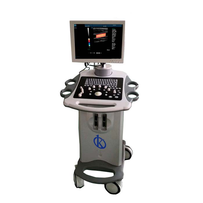
In this sense, ultrasound imaging is a valuable tool in medical diagnosis, but it has limitations. It cannot be used to image bones or other opaque structures and is less effective in obese patients. Despite this, at Kalstein we can offer you the ideal equipment with a large 15 inch color LED display, real Doppler function USB ports and VGA port and 2 probe connectors.
What is an Ultrasound?
Ultrasound is sound waves with frequencies higher than the upper audible limit of human hearing, not different from “normal” (audible) sound in its physical properties, except that humans cannot hear it. This limit varies from person to person and is about 20 kilohertz (20,000 hertz) in healthy young adults. Ultrasound devices operate at frequencies from 20 kHz to several gigahertz.
Medical ultrasonography (also known as diagnostic ultrasonography or ultrasonography) is an imaging technique based on the application of ultrasonography. It is widely used in various medical specialties, including cardiology, obstetrics and gynecology, urology, oncology, neurology and gastroenterology.
Ultrasound images are captured using a transducer that emits and detects ultrasound waves, and are transmitted through the body and bounce off organs, tissues, and fluids. Echoes are converted into electrical signals that are displayed on a computer screen as a real-time image.
Factors for Performing Ultrasound
Image quality depends on several factors, including the type of machine, the operator’s skill, and the type of fabric being created. Medical ultrasonography is generally considered safe when used for diagnostic purposes.
Some potential risks associated with ultrasound include:
- – Tissue warming (by absorption of acoustic energy).
- – Cavitation (formation and collapse of small bubbles in tissues).
- – Mechanical index (a measure of cavitation potential).
- – Thermal index (a measure of warming potential).
- – Peak-time spatial mean intensity (a measure of the maximum intensity of the ultrasound wave).
The American Institute of Ultrasound in Medicine (AIUM) has published safety guidelines that recommend limiting the thermal index and mechanical index to avoid potential damage.
Ultrasound is a valuable tool for diagnosing many different conditions. It can be used to visualize internal organs, blood flow, and fetuses during pregnancy. Ultrasonography can also be used to guide biopsies and other medical procedures.
Usage Applications
The use of ultrasound in medicine is constantly evolving, and new applications are being developed all the time. Some promising areas of research include:
- – Use ultrasound to noninvasively monitor the progression of Alzheimer disease and other forms of dementia.
- – Use ultrasound to speed healing of bone fractures.
- – Use ultrasound to noninvasively deliver drugs to specific areas of the body.
- – Use of ultrasound to improve the accuracy of cancer detection.
- – Use of ultrasound to improve the success rate of in vitro fertilization.
- – Using ultrasound to increase the effectiveness of physical therapy.
- – Use of ultrasound to treat baldness.
As medical ultrasound technology continues to improve, the potential uses of this modality will surely increase.
Portable Ultrasound Scanner Cart, Color Doppler Ultrasound Machine Brand Kalstein
We at Kalstein offer you the best medical equipment, with the most advanced technology on the market. We have for our customers the best Ultrasound Scanner, belonging to the YR series, has: Image modes: B 2B… 4B M B/C/B/D/B/C/D/B/CFM/D PDI. Dual Color Simultaneous 2D/. Composite color. Duplex/Triplex PW… CFM… CD Directional PD CDE. Among many other features you can see from our catalog HERE We are manufacturers and we have the best advice, so that your purchase is the ideal and at excellent prices. To do this, visit HERE