Curious fact
1671: Friar capuchino named Chérubin d’Orléans builds the microscope where you can see the reliefs of the samples in inverted form.
1871: Charles Wheatstone establishes the theoretical basis on how to build a stereoscopic or dissection microscope.
End of the 19th century: Horatio S. Greenough designed the first functional stereoscope and presents it to the German microscope manufacturer Carl Zeiss.
1957: a new type of stereoscopic or dissection microscope called CMO (Common Main Objective) was introduced.
Any kind of analysis equipment is a fundamental device to have in your laboratory. Yu must count with the highest quality equipment in order to achieve reliable results when performing tests. Laboratory microscopes are an important part of those devices since they are objects that help to gives better look at objects by magnifying them. Microscopes are to see objects that the naked eye cannot see. The name of the science that involves investigating small objects and structures is Microscopy.
Since there is a wide variety of microscope types, one way to group them is by the way the instruments interact with a sample in order to create images. That can happen by sending a beam of light, electrons to a sample in its optical bath or by scanning across and a short distance from the surface of a sample using a probe. Almost every industry now takes advantage of its capabilities.
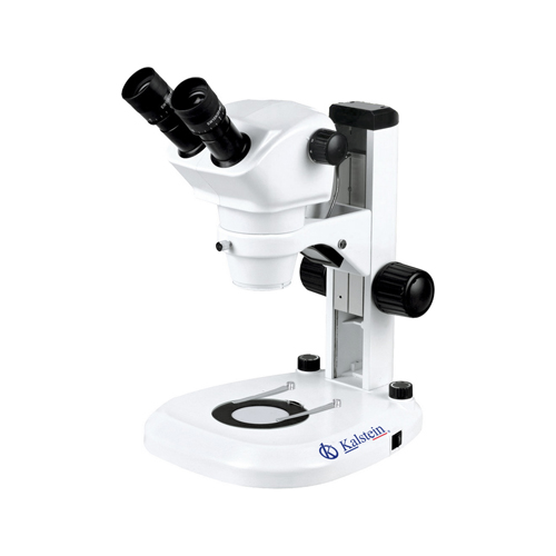
Clinical laboratory microscopes
You should get to know some of the technical details about the different microscope types. First, optical microscopes use light illumination, lenses and often a digital camera to magnify the images. In this microscope category, you can find stereomicroscopes and compound microscopes. The confocal and fluorescence microscopes permit live cell imaging by including laser scanning and spinning disk variations.
In the case of electron microscopes, these types use an electron beam to illuminate samples, this way it significantly enhances the resolving power. There is the Transmission electron microscopy (TEM), which has benefited from nowadays low-cost image processing and data storage. On the other hand, there is also the Scanning electron microscopy (SEM), which has the ability to give highly detailed depth of the examining field at a high magnification.
There is also the scanning probe microscopy, in which the images of a surface’s sample are produced. The scanning tunneling microscopy (STM) uses a stylus to produce detailed 3-D images. There is also atomic force microscopy (AFM) and near-field scanning optical microscopy, microscope types that fall under this heading.
High quality laboratory microscopes
It means a lot to us that you have the highest technology available in your laboratory. That is why we make a big effort to provide quality devices, especially when it comes to microscopes giving the huge importance nowadays. If you want to upgrade your instrumentation, or if you are staring a new laboratory, you should know that there are some key considerations to keep in mind when purchasing a laboratory microscope. You have to be aware that not all instrumentation perform in the same manner. In addition, imaging techniques can vary from system to system and these features make a big difference between one model and the other.
You must take into considerations the microscope type (decision that depends on your application field. In addition, you have to think about your laboratory space, especially the workspace, because you need to have comfort when using a microscope. Depending on your field or if you are using the microscope outside the laboratory because of an exploration field, you must think about portability.
As a conclusion, microscopy field is a very diverse one. Nowadays, not only biological laboratories need to have microscopes. Thanks to the technology advances and society’s needs, microscopy is an evolving market that is constantly evolving. Some of the fields you can now see them, are:
- Microscope attachments that permit nanomanipulation of individual cells and nanotubes.
- Techniques that enhance the tissue engineering process.
- New imaging methods such as multiphoton, TIRF, and super-resolution microscopy that have revolutionized cell biology.
In Kalstein Laboratory Equipment, we are offering you the high-end microscopes you need in your company. Visit our catalog HERE